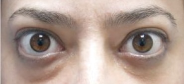
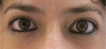
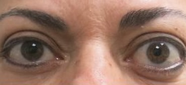
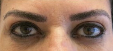
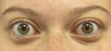
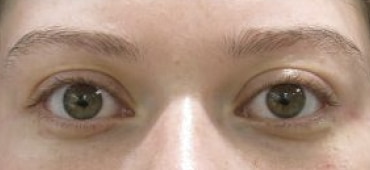
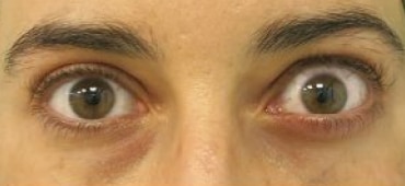
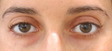
The cause of the disease is the infiltration of the orbital fat and eye muscles with inflammatory cells with exudate tissue edema. Fortunately, in many cases the orbital involvement is mild. The orbital disease is active for 6 to 12 months and then stabilizes, at which point corrective and reconstructive surgeries can be performed on the orbital and eyelids. In many cases, multiple surgeries are needed to restore the appearance of the eye and eyelids to the condition before the thyroid disease.